
When a person experiences persistent back pain, injuries, or discomfort that interferes with daily activities, a physician may recommend imaging studies to determine the source of the problem. However, many patients are unsure about the differences between an MRI, a CT scan, and X-rays.
Choosing the appropriate imaging study is essential for obtaining an accurate diagnosis and developing the most effective treatment plan. Each technology offers unique advantages and allows physicians to evaluate different structures within the body. In many cases, a Spine MRI plays a key role in identifying conditions that other imaging methods may not fully reveal.
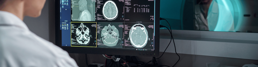
Why Are Imaging Studies Important for Back Pain?
Back pain can result from many different causes. Some are related to muscles and ligaments, while others involve intervertebral discs, nerves, vertebrae, or degenerative conditions.
Diagnostic imaging studies help physicians obtain detailed information about the affected structures and determine the underlying cause of symptoms more accurately.
Depending on the clinical situation, a healthcare provider may recommend one or multiple imaging tests, including a Spine MRI, CT scan, or X-ray.
What Do X-Rays Show?
X-rays are among the most commonly used diagnostic imaging tools in medicine. They are primarily used to evaluate bones and identify visible abnormalities in the vertebrae.
X-rays can help detect:
- Fractures
- Vertebral misalignment
- Advanced degenerative changes
- Structural spinal abnormalities
- Scoliosis
However, X-rays have significant limitations because they do not provide detailed visualization of soft tissues, intervertebral discs, nerves, or the spinal cord.
For this reason, X-rays are often used as an initial evaluation tool but may not provide enough information for a definitive diagnosis.
What Does a CT Scan Show?
Computed tomography (CT) uses X-ray technology combined with advanced computer processing to generate cross-sectional images of the body.
This study offers more detail than traditional X-rays and provides enhanced visualization of certain bony structures.
A CT scan may be useful for identifying:
- Complex fractures
- Traumatic injuries
- Bone abnormalities
- Structural changes within the spine
However, when the goal is to evaluate discs, nerves, the spinal cord, or soft tissues, a Spine MRI often provides significantly more detailed information.
What Does a Spine MRI Show?
A Spine MRI is considered one of the most comprehensive imaging studies available for evaluating back pain and spinal conditions.
Unlike X-rays and CT scans, a Spine MRI uses magnetic fields and radio waves to create highly detailed images without exposing patients to ionizing radiation.
This technology allows physicians to visualize:
- Intervertebral discs
- Spinal nerves
- The spinal cord
- Muscles
- Ligaments
- Inflammation
- Herniated discs
- Nerve compression
A Spine MRI is frequently recommended when patients experience neurological symptoms, persistent pain, or conditions that cannot be adequately assessed through other imaging methods.
Which Study Is Best?
There is no single imaging study that is ideal for every patient. The most appropriate choice depends on symptoms, medical history, and the specific information needed for diagnosis.
X-Rays
Best for evaluating basic bone structures and identifying fractures or deformities.
CT Scan
Useful for traumatic injuries and detailed assessment of bone tissue.
Spine MRI
A Spine MRI is widely regarded as one of the most valuable imaging studies when physicians need to examine soft tissues, nerves, discs, and internal spinal structures in detail.
Because of its ability to provide highly detailed images, many specialists consider a Spine MRI the preferred option for investigating complex or persistent spinal symptoms.
When Should You Seek Medical Evaluation?
You should consult a healthcare professional if your pain:
- Persists for several weeks
- Becomes increasingly severe
- Limits daily activities
- Is accompanied by tingling or weakness
- Radiates into the arms or legs
- Develops after an injury or accident
Timely diagnosis can help prevent complications and support more effective treatment outcomes.
Convenient Access for Patients on Both Sides of the Border
Location can be an important factor when seeking specialized diagnostic imaging. Diagnóstico de Las Américas is located just minutes from the Bridge of the Americas, providing convenient access for patients from Ciudad Juárez, El Paso, and surrounding border communities.
With advanced imaging equipment and experienced healthcare professionals, we provide high-quality diagnostic services, including Spine MRI studies, designed to support accurate and reliable diagnoses.
Conclusion
X-rays, CT scans, and MRI examinations each serve different purposes in medical diagnosis. The best option depends on the individual patient, their symptoms, and the clinical information required by the physician.
If you are experiencing persistent back pain or symptoms that affect your quality of life, consulting a medical professional and obtaining the appropriate imaging study may be the first step toward identifying the cause and beginning effective treatment. In many cases, a Spine MRI can provide the detailed information needed to guide medical decision-making and improve patient outcomes.